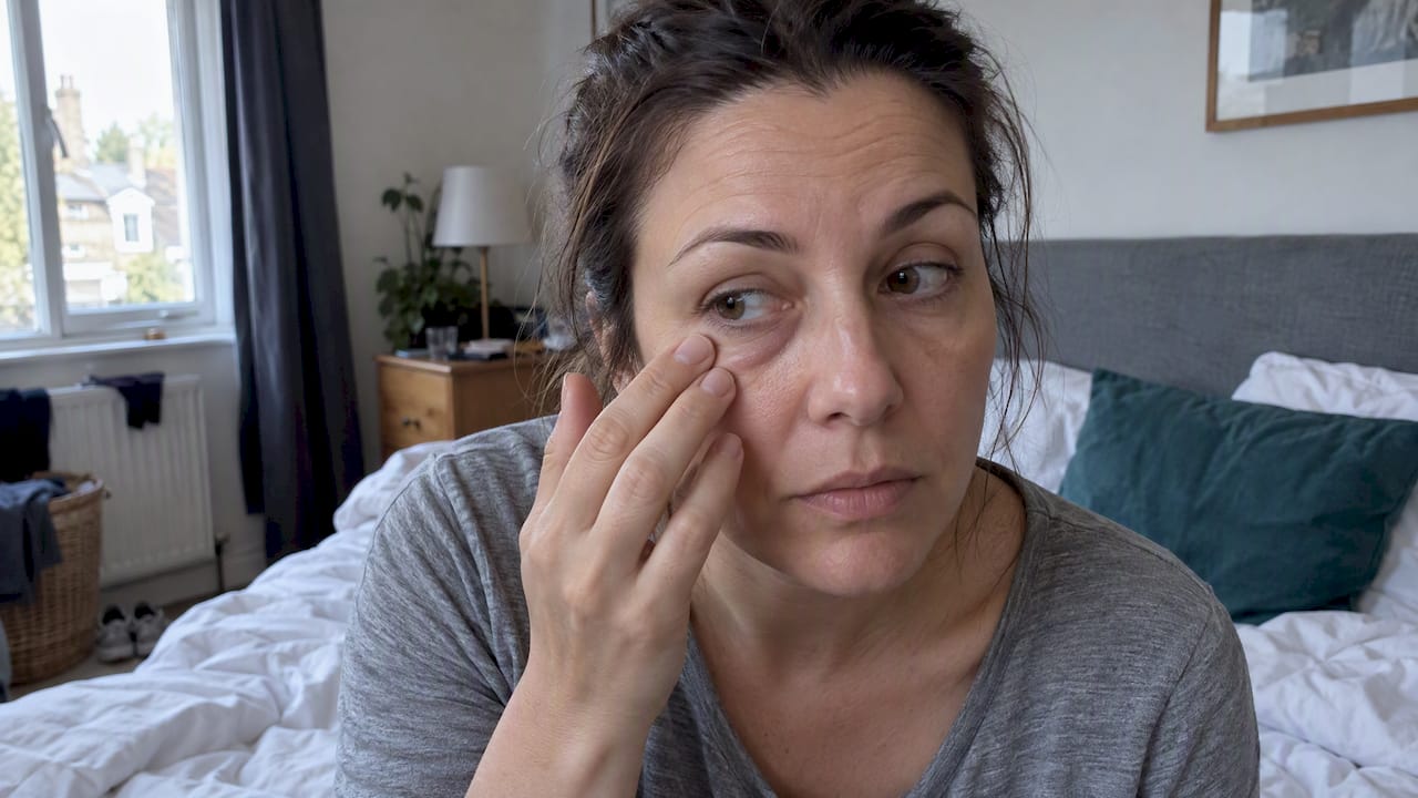
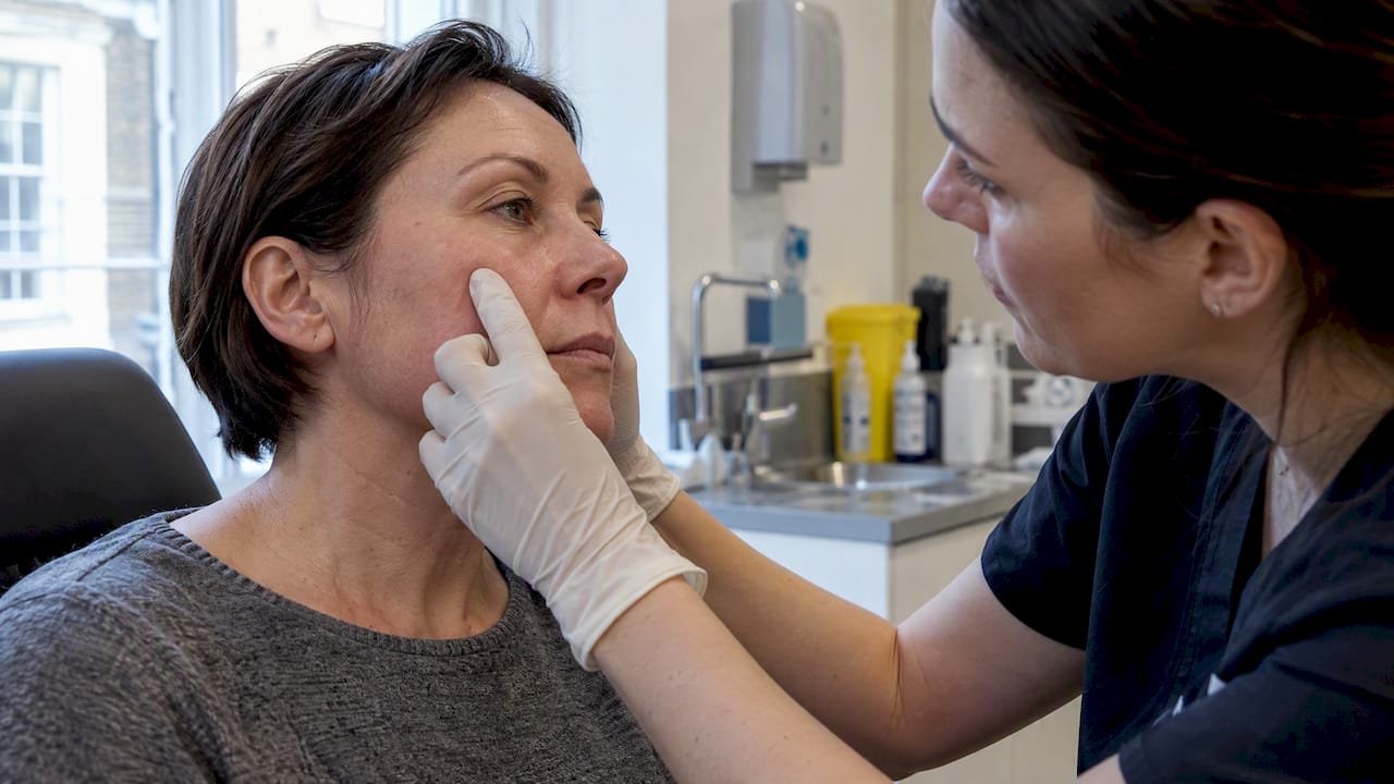

If you notice blanching or pallor, pain that feels disproportionate or keeps building, coolness in the treated area, or slow capillary refill after a filler treatment, treat this as a possible vascular occlusion and seek urgent assessment straight away. Occlusion is rare, but outcomes depend heavily on speed: earlier treatment limits tissue damage, which is why bodies like the ACE Group treat it as a medical emergency with a clear response protocol.
TL;DR:
- A normal capillary refill takes one to two seconds; delayed return, blanching, coolness, or worsening pain can signal occlusion, even days after treatment.
- Unlike bruising, which fades gradually and does not blanch under pressure, occlusion can progress from pallor to mottling; local anesthetic may hide early pain.
- Call the treating clinic immediately, take photos, and seek emergency care if symptoms are severe or prompt assessment is unavailable; do not massage the area.
- For hyaluronic acid filler occlusion, clinicians use hyaluronidase across the affected area, often in repeated doses; treatment within 48 hours lowers necrosis risk.
- Before booking, confirm the clinic stocks hyaluronidase and has trained staff plus an emergency protocol; conservative injections reduce risk in vulnerable areas.
Table of Contents
- Signs of vascular occlusion and when they typically show up
- How to tell vascular occlusion apart from normal bruising
- What to do immediately if you suspect an occlusion
- What treatment for vascular occlusion actually involves
- Follow-up, monitoring and what should prompt a return visit
- Reducing your risk and choosing a clinic prepared for emergencies
- How common occlusion is and why timing shapes the outcome
- Why occlusion can appear late and what delayed signs look like
- Our perspective on vigilance and safety
- FAQ
- Sources
Signs of vascular occlusion and when they typically show up
Vascular occlusion happens when filler blocks or compresses a blood vessel, cutting off circulation to the surrounding tissue. The clinical picture is distinctive once you know what to look for, and it rarely looks like an ordinary bruise.
The main red flags are:
- Immediate pallor or blanching: the skin turns strikingly pale or white, often in a defined pattern rather than a general blotch.
- Severe or escalating pain: discomfort that is far greater than you would expect from an injection, or that keeps intensifying rather than settling.
- Livedo reticularis: a lace-like, mottled or reticulated discolouration across the skin.
- Dusky, blue or grey tones: a sign that tissue is being starved of oxygen.
- Coolness: the affected area feels noticeably colder than the surrounding skin.
- Delayed capillary refill: pressing the skin and releasing it shows a slow return of colour.
According to Cleveland Clinic, typical symptoms include pain, swelling and progressive changes in skin colour, and early diagnosis gives the best chance of full recovery. Most of these signs appear immediately or shortly after injection, though delayed presentations can occur.
Occlusion is estimated to affect around 0.001% of filler treatments, according to guideline data published in PMC, which also tracks the typical progression from pallor through livedo reticularis to potential necrosis if untreated.
Compare this with the reactions we expect after most filler appointments: redness, mild swelling and bruising. These are normal, settle within a few days and usually clear within about a week, according to guidance from the Pan London Facial Lipoatrophy Service. The distinction matters because bruising stays put and fades gradually, while occlusion signs tend to appear fast and get worse rather than better.
How to tell vascular occlusion apart from normal bruising
The clearest distinguishing feature is blanching. A bruise is caused by blood leaking under the skin, so it does not turn white when pressed, it stays discoloured. Vascular compromise behaves differently: the skin often blanches first, then progresses to the reticulated, net-like mottling of livedo reticularis as circulation struggles to reach the tissue.
Capillary refill time (CRT) is the most practical test you or a clinician can use to check this:
- Press firmly on the affected skin for a few seconds.
- Release the pressure and watch how quickly colour returns.
- A clinician can assess how quickly colour returns after pressure is released.
- A delayed return or other concerning skin changes warrant urgent clinical assessment.
- A brisk refill paired with bluish discolouration can instead point to venous compression, according to PMC guidance on hyaluronic acid filler-induced vascular occlusion.
Pain patterns matter too, though they can be confusing immediately after treatment. Local anaesthetic used during injection can mask early pain signals, so the absence of severe pain in the first few minutes does not rule out occlusion. If pain develops or worsens once the anaesthetic wears off, that is still significant.
Pro Tip: If you are unsure whether what you are seeing is a bruise or something more serious, contact your clinic rather than waiting to see if it resolves on its own; a quick check costs nothing and delay is the one thing that makes outcomes worse.
Skin tone and pre-existing circulation issues can change how blanching and discolouration present. Pallor can be harder to spot on darker skin tones, and people with conditions affecting peripheral circulation may show different baseline refill times. None of this is something to self-diagnose with confidence, which is exactly why the threshold for contacting a clinic should stay low whenever something looks or feels wrong.
What to do immediately if you suspect an occlusion
Acting within minutes, not hours, is the single factor most within your control. Follow this sequence:
- Contact your treating clinic immediately, by phone if possible, and describe your symptoms clearly.
- If the clinic cannot assess you promptly, or your symptoms are severe (rapidly spreading discolouration, severe pain, visible skin changes), go to emergency care without waiting for a callback.
- Take photos of the affected area as symptoms develop, as this helps the clinician assess progression even before you arrive.
- Avoid massaging or applying pressure yourself unless a clinician specifically instructs you to, since inappropriate manipulation can worsen tissue compromise.
- Attend in person as soon as you can, bringing details of exactly when and where you were injected.
When you contact the clinic, be ready to share:
- The exact time and location of your injection.
- A clear description of your symptoms and when they started.
- Photos showing colour and texture changes.
- Any known allergies, particularly to local anaesthetics or hyaluronidase.
- Current medications, including blood thinners.
A well-prepared clinic can assess the skin and circulation and decide whether urgent treatment is needed. Follow-up should be guided by the treating clinician. Our own guidance on filler side effects and when to get same-day care covers this same escalation pathway in more detail.
What treatment for vascular occlusion actually involves
Hyaluronidase is the principal treatment for occlusion caused by hyaluronic acid filler, and timing drives the outcome. According to PMC management guidance, the standard pathway once occlusion is suspected is to stop injecting immediately, assess capillary refill and skin appearance, then infiltrate hyaluronidase across the entire compromised area rather than a single point.
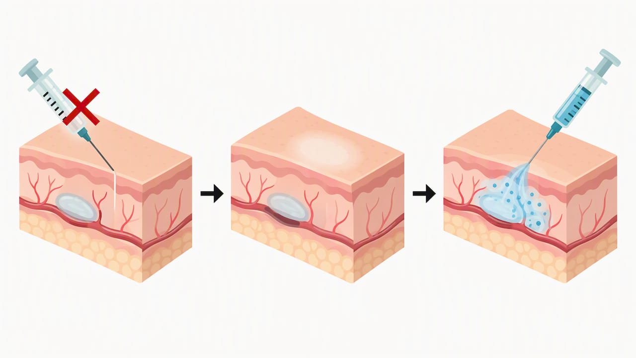
Treatment and follow-up should be managed by a qualified clinician, who can assess the affected area and decide what care is needed.
The ACE Group frames this as a high-dose, pulsed protocol: rather than a single conservative dose, clinicians give repeated, substantial doses hourly until clinical signs show blood flow has returned. This aggressive approach reflects how time-sensitive tissue viability becomes once a vessel is compromised.
Guideline data shows that treatment within 48 hours meaningfully reduces the risk of tissue necrosis, according to the same PMC guidelines, which is the clinical basis for treating every suspected occlusion as urgent rather than waiting to see how it develops.
Beyond the enzyme itself, a thorough response includes observing the patient for any allergic reaction to hyaluronidase, providing soothing topical aftercare if any skin breakdown has occurred, and referring to a specialist if complications are severe or slow to resolve. Clinics managing this well should give you written aftercare instructions and check in daily until the issue is fully resolved, rather than leaving you to monitor yourself after the initial visit.
Follow-up, monitoring and what should prompt a return visit
Once treatment has started, monitoring does not stop at the first dose of hyaluronidase. Clinics managing an occlusion properly will want to see you daily until the skin has visibly stabilised, often photographing the area at each visit to track whether colour and texture are genuinely improving rather than just appearing to.
Signs that mean you should go back immediately, even after treatment has begun, include:
- Spreading discolouration rather than a shrinking, improving area.
- Blistering or any new skin breakdown.
- Increasing pain, rather than gradual easing.
- Signs of infection: warmth, pus, or a fever.
Observation after hyaluronidase is also about more than the occlusion itself. Allergic reactions, while uncommon, can occur, so clinics typically watch patients for a period after infiltration. Basic wound care, such as keeping any broken skin clean and protected, supports healing and reduces the chance of secondary infection while the tissue recovers.
Reducing your risk and choosing a clinic prepared for emergencies
Most of the risk reduction here happens before you are ever injected, through technique and clinic choice rather than anything you do afterwards.
- Conservative injection technique in high-risk zones (such as the glabella, nose and tear trough) lowers the chance of vessel compromise.
- Lower injection volumes per pass reduce pressure build-up around vessels.
- An experienced injector who understands facial vascular anatomy is better placed to avoid danger zones.
- Proper informed consent, including a clear explanation of what occlusion looks like, means you know what to watch for afterwards.
When choosing where to be treated, check that the clinic has immediate access to hyaluronidase, trained staff who can recognise and manage occlusion, and formal emergency protocols rather than an improvised response. CQC registration and membership of a recognised safety body such as the ACE Group are practical ways to confirm this standard. The MHRA and UK government guidance is explicit that public safety depends on treatments being carried out by properly trained, registered professionals with emergency medicines on hand, since botched procedures can cause serious harm including necrosis and, in rare cases, blindness.
Pro Tip: Before booking, ask directly whether the clinic stocks hyaluronidase on-site and what their response time looks like if you call with a concern; a confident, specific answer tells you more than any marketing claim.
Our guide to choosing a dermal filler clinic and our explainer on why registered practitioners matter both go through this checklist in more depth.
How common occlusion is and why timing shapes the outcome
Vascular occlusion is rare. Guideline estimates put the incidence of impending necrosis after dermal filler at around 0.001%, or roughly one case in every 100,000 treatments, according to PMC guideline data, though reporting limitations mean the true figure could differ slightly from registry to registry.
What matters more than the rarity is the timing of treatment once it occurs. Prompt treatment may reduce the risk of tissue damage, so suspected occlusion needs urgent clinical assessment. Severe outcomes such as necrosis and, in extremely rare cases, blindness have been reported, which is precisely why speed, not alarm, is the appropriate response to any suspected occlusion.
Why occlusion can appear late and what delayed signs look like
Not every occlusion announces itself straight away. Delayed presentation can happen when an embolus lodges at a vessel branch point and gradually restricts flow, or when swelling after injection compresses a vein over the following hours.
Watch for these delayed signs over the days after treatment:
- Progressive livedo reticularis that spreads rather than fades.
- Blistering or small pustules erupting on the skin.
- Darkening or sloughing of the skin surface.
Any of these signs, even days after a seemingly uneventful treatment, warrant the same urgent response as symptoms appearing immediately: contact your clinic and seek assessment without delay.
Our perspective on vigilance and safety
Most filler treatments pass without complication when performed by a properly trained practitioner who understands facial anatomy and injects conservatively in high-risk areas. The rare cases that do occur reinforce something we believe strongly: the value of a clinic isn’t only in the treatment itself, it’s in how quickly and competently it responds when something doesn’t go to plan.
We hold CQC registration and ACE Group membership. If you notice any of the signs covered here after treatment with us or elsewhere, contact a clinic immediately rather than waiting to see how things develop.
— Vishul
This article is general information, not a substitute for advice from a qualified doctor. Consult a qualified healthcare professional about your own circumstances before acting on anything here.
FAQ
How long after fillers does vascular occlusion happen?
Most cases appear immediately or within the first few hours after injection, though rare delayed presentations can develop over the following hours to days, according to PMC guidance on hyaluronic acid filler complications. Any new or worsening symptoms during this window should prompt urgent clinic contact.
What does an occlusion after lip fillers look like?
An occlusion typically shows as pallor or blanching of the skin, pain that feels disproportionate to the injection, and sometimes a lace-like mottled pattern known as livedo reticularis. The area may also feel cool to the touch, which distinguishes it from the warmth of ordinary post-treatment swelling.
How do you know if you have an occlusion?
The clearest bedside check is capillary refill time: press the skin and watch how quickly colour returns, with a normal refill taking one to two seconds according to PMC clinical guidance. A delayed return, combined with pallor, disproportionate pain or coolness, points toward occlusion and needs urgent assessment rather than a wait-and-see approach.
Do vascular occlusions always hurt?
Not always, and this is part of what makes them tricky to spot. Local anaesthetic used during injection can mask early pain, and according to Cleveland Clinic, pain is one of several possible symptoms alongside swelling and changes in skin colour, so visual signs matter just as much as discomfort.
Sources
- This month’s guideline: The Use of Hyaluronidase in Aesthetic Practice (v2.4) – PMC
- What Is a Vascular Occlusion?