
TL;DR:
- D imaging generates interactive, patient-specific models that enhance communication and support accurate treatment planning.
- This technology improves patient understanding through visual simulation, reducing anxiety, and enabling real-time dialogue.
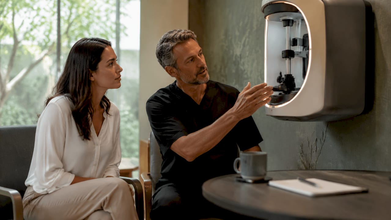
3D imaging in cosmetic consultations is defined as a technology that generates interactive, patient-specific three-dimensional models of the face or body to support clinical planning and shared decision-making. The standard industry term for the most widely used capture method is stereophotogrammetry, though the broader category is often called 3D surface imaging. Systems such as Vectra 3D Imaging have made this technology a practical fixture in aesthetic clinics, replacing flat photographs with volumetric views that both patient and clinician can rotate, annotate, and discuss in real time. The role of 3D imaging in consultation goes well beyond visual novelty. It is a communication and planning tool that aligns expectations, supports informed consent, and gives clinicians objective anatomical data before any treatment begins.
How does 3d imaging improve patient understanding?
3D imaging replaces abstract verbal descriptions with something you can see and interact with. When a clinician explains that dermal filler will restore volume to the mid-face, the words alone rarely convey what that means for your specific face. A rotatable 3D model does.
The benefits for patient education are well documented. Multimedia visual aids during the consent process improve patient knowledge compared to brochures alone, according to a 2025 clinical study. This matters because aesthetic procedures have subjective outcomes, and a patient who genuinely understands what is planned is far better placed to give meaningful consent.
Interactive viewing also creates space for real dialogue. You can point to areas of concern on the model, ask the clinician to show you a simulated change from a different angle, and leave the consultation with a clear visual memory of the discussion. That kind of engagement is simply not possible with a 2D photograph.
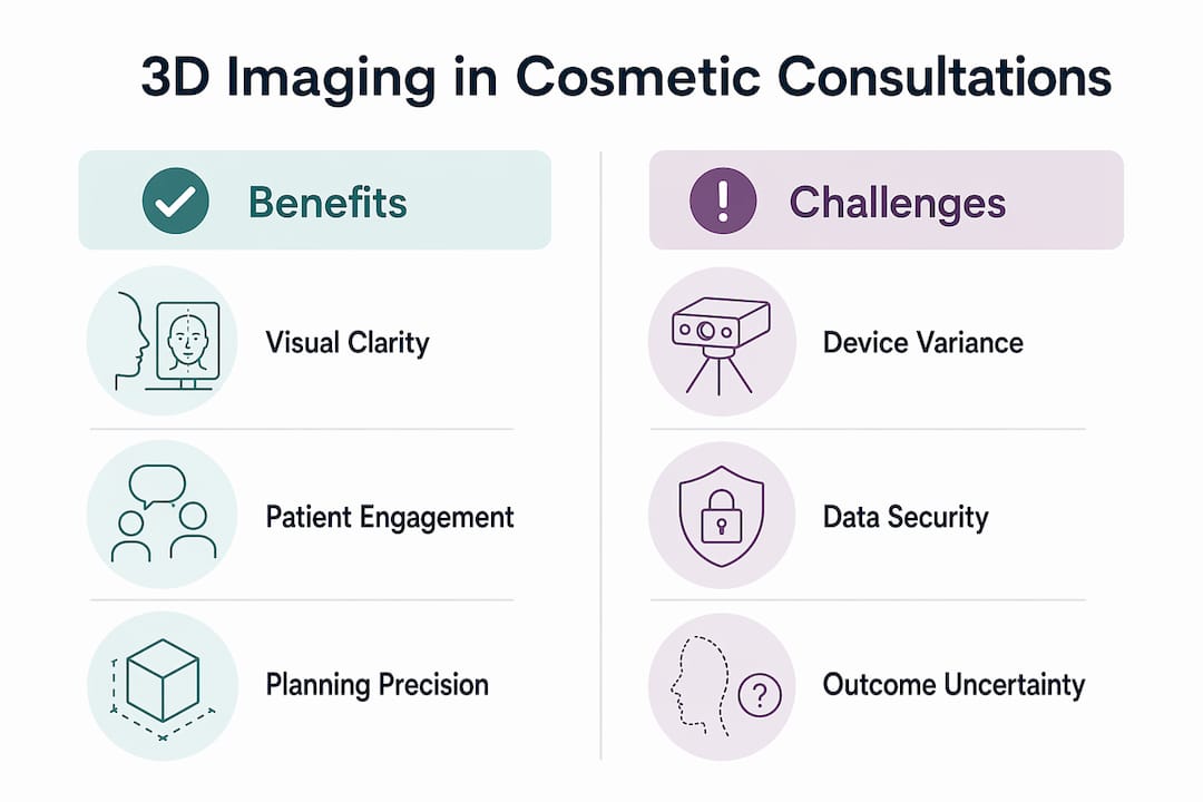
Key benefits of 3D imaging for patient understanding include:
- Side-by-side simulation views showing pre-treatment anatomy alongside a proposed outcome
- Multi-angle rotation so you can assess changes from the front, side, and three-quarter positions
- Real-time feedback allowing you to request adjustments to the simulation during the appointment
- Reduced anxiety because visual transparency of limitations and outcomes builds trust in the clinical plan
Pro Tip: Always ask your clinician to clarify that a 3D simulation shows a possible outcome, not a guaranteed result. Simulations are planning tools, not promises, and understanding this distinction protects you from unrealistic expectations.
How does 3d imaging support personalised treatment planning?
3D imaging gives clinicians objective, measurable data about your anatomy that no 2D photograph can provide. Stereophotogrammetry is used in the majority of clinical studies on facial scanning, with applications spanning surgical planning, symmetry analysis, and outcome monitoring. The technology is more reliable and reproducible than conventional 2D assessment, which means treatment decisions rest on firmer ground.
Here is how 3D imaging supports the planning process step by step:
- Anatomy capture A full facial scan creates a precise digital model of your soft-tissue contours, proportions, and asymmetries before any discussion of treatment begins.
- Symmetry analysis The clinician uses the model to identify and quantify asymmetries, which informs decisions about filler placement or Botox injection patterns.
- Outcome visualisation Simulated changes are applied to the model so both parties can review the proposed result before committing to a plan.
- Baseline documentation The pre-treatment model serves as a precise, storable record for clinical follow-up and medicolegal purposes.
- Workflow integration 3D imaging is not limited to the consultation room. It supports preoperative to postoperative stages across the full treatment workflow.
| Feature | 2D Photography | 3D Surface Imaging |
|---|---|---|
| Depth and volume data | No | Yes |
| Reproducibility | Variable | High |
| Symmetry measurement | Subjective | Objective |
| Interactive simulation | No | Yes |
| Medicolegal documentation | Limited | Detailed |
One important caveat: device and software heterogeneity across the industry means that results from one system are not always directly comparable to another. Clinicians must account for this variation when presenting simulations and avoid framing any outcome as absolute.
What are the technical challenges of 3d imaging in consultations?
3D imaging is a powerful tool, but it is not without practical limitations. Understanding these challenges helps you ask better questions during your consultation and interpret what you see on screen with appropriate context.
Accuracy is the primary concern. Patient movement during capture causes soft-tissue distortions that reduce the reliability of the resulting model. Even a slight shift in expression or head position between the camera’s multiple capture points can introduce artefacts. Clinics that follow strict positioning protocols and take multiple captures reduce this risk significantly.
The main technical and practical challenges include:
- Motion artefacts caused by patient movement during the scan, which distort soft-tissue geometry
- Software heterogeneity across different 3D imaging platforms, making standardised comparisons difficult
- Capture protocol variation between clinics, which affects the consistency of results
- Data security obligations requiring that 3D facial models are stored securely and handled under clear patient consent
- Clinician training requirements because the value of a scan depends heavily on the skill of the person interpreting and presenting it
Standardisation of scanning devices and post-processing protocols is an active area of development in the field. Until universal standards exist, the quality of your 3D consultation experience will vary between clinics.
Pro Tip: Before your consultation, ask the clinic which 3D imaging system they use and how they store your facial data. A reputable clinic will have a clear data consent process and will explain the limitations of their specific system before showing you any simulation.
How does 3d imaging transform the consultation experience?
The consultation itself changes fundamentally when 3D imaging is introduced. 3D imaging shifts the workflow by front-loading the anatomy capture before in-depth verbal discussion begins. This means the clinician already has your facial model on screen when the conversation about treatment starts, enabling real-time interactive simulation throughout the entire appointment rather than at the end.

This image-first approach has a measurable effect on the quality of the dialogue. Instead of the clinician describing what they plan to do while you try to visualise it, you are both looking at the same reference point. Goals, limitations, and alternatives are discussed in relation to a shared visual object. That shift from verbal-first to image-first consultation is one of the most significant practical advantages of 3D imaging in aesthetic practice.
The table below compares a traditional consultation with one enhanced by 3D imaging:
| Consultation Feature | Traditional Approach | 3D Imaging-Enhanced Approach |
|---|---|---|
| Visual reference | 2D photographs or verbal description | Interactive 3D model of your face |
| Expectation setting | Clinician-led verbal explanation | Shared simulation reviewed together |
| Patient engagement | Passive listening | Active participation and feedback |
| Anatomical precision | Subjective assessment | Objective measurement and symmetry data |
| Consent quality | Brochure or verbal summary | Visual, documented, interactive process |
| Follow-up comparison | Before-and-after photos | Precise 3D baseline versus outcome model |
The emotional impact of this shift is also worth noting. Seeing a simulation of a proposed change reduces anxiety because it replaces uncertainty with a concrete visual reference. You are not imagining what you might look like. You are reviewing a model and deciding whether the direction feels right. That collaborative dynamic builds trust and makes the consent process more meaningful. For procedures such as facial anatomy assessment, this level of precision is particularly valuable.
The role of consultation in aesthetics has always been about aligning clinical expertise with patient goals. 3D imaging gives both parties a shared language to do that more effectively.
Key takeaways
3D imaging in cosmetic consultations is most valuable as a shared communication tool that improves expectation alignment, supports precise planning, and strengthens the informed consent process.
| Point | Details |
|---|---|
| Communication over prediction | 3D simulations are planning aids, not guaranteed outcomes; always clarify this with your clinician. |
| Reproducible anatomical data | Stereophotogrammetry provides more reliable soft-tissue assessment than 2D photography for treatment planning. |
| Improved consent quality | Multimedia and 3D visual aids significantly improve patient knowledge compared to brochures alone. |
| Technical limitations exist | Motion artefacts and software variation affect accuracy; strict protocols and clinician skill are critical. |
| Workflow transformation | Image-first consultations enable real-time simulation throughout the appointment, improving collaboration. |
Why 3d imaging is a communication tool, not a crystal ball
From my experience working in aesthetic medicine, the single biggest misunderstanding about 3D imaging is that patients sometimes arrive expecting a guarantee. They have seen the simulation, they have approved the direction, and they believe the result is contractually fixed. It is not. The simulation is a map, not a destination.
What 3D imaging does exceptionally well is give both the clinician and the patient a shared visual vocabulary. Before this technology, a clinician might spend twenty minutes trying to explain why a small amount of hyaluronic acid filler placed in the tear trough would change the perception of the entire mid-face. Now, you show it. The conversation becomes faster, more precise, and far more honest about what is and is not achievable.
The technology also forces clinicians to be more rigorous. When you are building a 3D model and presenting a simulation, you cannot be vague. You have to commit to a visual direction, which means you have to think carefully about anatomy, proportions, and the patient’s specific goals before the appointment even begins. That discipline benefits everyone.
My honest view is that the clinics not yet using 3D imaging are leaving a significant communication gap in their consultations. The technology is not perfect, and standardisation across devices remains an ongoing challenge. But the direction of travel is clear. 3D imaging will become the baseline expectation for any serious aesthetic consultation, not a premium add-on.
— Vishul
Experience 3d imaging at Theaestheticsroom
Theaestheticsroom integrates advanced 3D imaging technology into consultations at our Knightsbridge clinic, giving you a precise visual foundation for every treatment decision. Whether you are exploring Botox treatments or considering dermal fillers, your consultation begins with a detailed anatomical assessment that supports personalised planning and clear expectation management.
Our CQC-accredited practitioners use 3D visualisation to walk you through proposed changes in real time, so you leave your consultation with clarity and confidence rather than uncertainty. Book your consultation at Theaestheticsroom today and experience the difference that precise, image-led planning makes to your aesthetic journey.
FAQ
What is the role of 3d imaging in a cosmetic consultation?
3D imaging creates an interactive, patient-specific model of your face or body that clinicians use to plan treatments, visualise outcomes, and support informed consent. It replaces verbal descriptions with a shared visual reference that both you and your clinician can review together.
Is a 3d simulation a guarantee of my results?
No. 3D simulations are educational planning tools that show a possible direction, not a guaranteed outcome. Clinicians present them to improve communication and expectation alignment, not to make promises about final results.
How accurate is 3d facial scanning?
Accuracy depends on the system used and the capture protocol followed. Motion artefacts and soft-tissue distortion can reduce reliability, which is why strict positioning protocols and multiple captures are recommended for consistent results.
What happens to my 3d facial data after the consultation?
Your 3D model should be stored securely under a clear data consent agreement. Reputable clinics treat these digital records as medicolegal documents and will explain their data handling policy before capturing any images.
Does 3d imaging improve the informed consent process?
Yes. Research shows that multimedia visual aids during consent significantly improve patient knowledge compared to brochures or verbal explanations alone, making 3D imaging a meaningful upgrade to the standard consent process.